THE SCIENTIFIC FACTS ON
THE HOLY SHROUD
·
 Why is the Holy Shroud a scientific
object?
Why is the Holy Shroud a scientific
object?
·
What is the Shroud?
·
The Shroud’s physical characteristics
·
The markings on the Shroud
·
The Image on the Shroud
·
The wound and blood markings on the Shroud
·
Other substances found on the Shroud
·
Images of Roman coins from A.D. 29 over
the eyes of the Man in the Shroud
·
The three-dimensional nature of the image
on the Shroud
·
The Shroud image examined by physics and chemistry
·
The chemical nature of the image
·
How did a chemically-produced image get onto
the Shroud from an enveloped dead body?
·
Fraud Theories of Image formation
The painting
fraud theory
The Renaissance ‘photographic
genius’ theory
The ‘hot statue scorch’ theory
·
Scientific Theories of Image formation
Was the Image made by perspiration,
embalming spices, and vapours?
Was the Image made by radiation?
·
The attenuation of the radiation that is
required
·
The energy needed for radiation to produce
the image. Microwave radiation and/or proton flux
·
Conclusions from the Scientific Facts
·
Where could the (microwave/proton) radiation
come from?
·
A new binary, world view needed?
Variable speed of light (VSL) theories ? ‘Dark matter and dark
energy’?
Why is the Holy Shroud a Scientific Object?
The study of the Shroud
involves physics and chemistry because the Shroud is a linen cloth, and, as a
physical object, it is subject to scientific, quantitative examination and
evaluation. Of course, the Shroud is also of intense interest in many fields of study, such as forensic medicine,
art, iconography, archaeology, numismatics, textile science, history, theology
and devotional studies, and each of these disciplines has its own methods and
standards. If we concentrate here primarily on the physical and chemical
aspects of the Holy Shroud, this will in no way question the validity of the
other fields of study.
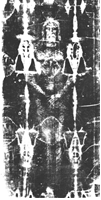
What is the Shroud? The Shroud is an ancient piece of yellowed
or ivory-coloured linen now in the Royal Palace Chapel in
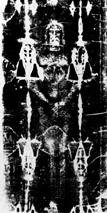
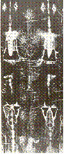
On one side of the cloth only, the Shroud
bears the front and back, full length, negative image of the naked body of a dead, scourged
crucified man about 5 ft 9 inches tall
and weighing about 175 pounds (75 kg.) .
The wound marks to the body, wrists, ankles and head match the wounds reported
in the four gospels of the New Testament as having been inflicted on Jesus
Christ during his crucifixion.
THE PHYSICAL CHARACTERISTICS AND MARKINGS
The Shroud’s Physical Characteristics. The cloth is a fine linen with a
three-to-one herringbone weave. The colour is a very pale ivory-yellow, consistent with its
reputed age and history. A detailed discussion of the cloth as a textile is
given by
Each thread of the linen consist of a
bundle of 80 -100 tiny linen fibrils about
one-tenth the thickness of a human hair (i.e. 15 -20 microns, 0.015 -0.020 mm). Linen is nearly
100% cellulose. Cellulose is polycellobiose with the chemical formula
(C6H10O5)x .

Linen
thread Individual
fibril
 The
Shroud is 436 cm long by 110 cm wide.
Sewn along one long side of the Shroud
is a ‘side strip’ about 8 cm wide of the same linen as the main Shroud,
the function of which is obscure, but which may
have been added to make the image centred or balanced on the Shroud for
ceremonial display purposes.(The Side Strip )
The
Shroud is 436 cm long by 110 cm wide.
Sewn along one long side of the Shroud
is a ‘side strip’ about 8 cm wide of the same linen as the main Shroud,
the function of which is obscure, but which may
have been added to make the image centred or balanced on the Shroud for
ceremonial display purposes.(The Side Strip )
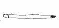
There are 22 triangular patches sewn into
the Shroud to cover holes in the Shroud made in a fire in 1532 which took place
in the Sainte Chapelle of the castle of Chambery in the French Alps, where the
Shroud was kept by the Dukes of Savoy from its acquisition by them from the de
Charny family in 1460 A.D. until its
final transfer to their Royal Palace in Turin in 1578 A.D. There is a backing cloth - the so-called
The specific
weight, unit weight or area-density of the linen, ( that is the weight per unit
area ) is about 23 milligrams per
square centimeter [3], so that the Shroud as a whole would weigh 436 x 110 x 23
x 10-6 = 1.1 kilograms. (This ‘specific weight’ or ‘unit weight’
measurement is an important one in calculating the possible contamination of the radiocarbon dating
samples cut in 1988 from a heavily
handled corner of the Shroud, since these samples averaged 42.9 mg/sq.cm, in weight, instead
of the 23 mg/sq. cm. for the rest of the Shroud, thereby showing a contamination of the test samples
of about 87% ( 42.9/23) = 1.87 (See Carbon 14 dating ).
The. physical qualities of the linen and
the image it bears have been studied
optically, by spectral reflectance, infrared and X ray reflectance, X-ray
fluorescence, and by many other scientific techniques [4,5,6,7,8,9].
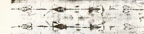
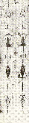
The Shroud’s Markings
There are two main types of markings on
the Shroud, namely the negative image of
the body, and many bloodstains. There are also a number of water stains
on the cloth made by the water used to douse the 1532 fire at
For detailed,
high quality photographs of the Shroud’s images and markings see various monographs such as that of Moretto
[10 ] and various Websites as follows:
Official Shroud of Turin Site in
The Holy Shroud Guild,
Collegamento pro Sindone Site,
The Shroud of
The Shroud of Turin, USA ( www.shroudofjesus.com/ )
(Other websites are to be found in various
search engines such as Google,
Scirus, AltaVista, etc.
The
Image Markings on the Holy Shroud
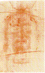
As seen by the naked eye, and in a
photographic positive, the image of the body of The Man in the Shroud appears
as a faint, pale brownish, or pale sepia coloring on the
ivory-yellow linen cloth. This image is on one side only of the cloth;
it shows the full length, front and back image of a dead man, as though the corpus
had been laid out on the extended
cloth, which was then folded back over the head to fully envelope or enshroud
it in the tomb. The image has a negative
character, since prominent body features such as the forehead, tip of the nose,
chin, chest, forearms, hands and knees, which would be closest to the
enveloping shroud, show up as the darkest shadings of sepia, while recessed
body areas such the eye sockets, neck, pelvis, and ankles are lightest.
 This
negative aspect was first discovered when Secundo Pia took the first photograph
of the Shroud in 1898. To his amazement,
the photographic negative came out of the developing bath as a positive image.
This astounded the scientific world of
the time and launched the scientific and scholarly efforts of the past century.
The hair and beard are visible as well as the flesh areas of the body. There
are faint indications that some deeper parts of the body are imprinted in the
image, such as the bones of the fingers and the teeth sockets, in a manner somewhat
reminiscent of an X-ray.
This
negative aspect was first discovered when Secundo Pia took the first photograph
of the Shroud in 1898. To his amazement,
the photographic negative came out of the developing bath as a positive image.
This astounded the scientific world of
the time and launched the scientific and scholarly efforts of the past century.
The hair and beard are visible as well as the flesh areas of the body. There
are faint indications that some deeper parts of the body are imprinted in the
image, such as the bones of the fingers and the teeth sockets, in a manner somewhat
reminiscent of an X-ray.
The image is sharply truncated or cut off
at the sides of the face and body. That is to say, if the image is envisaged as
having been formed by a stream of light photons, or other radiation, then the
radiation must have been strictly up and down, parallel to the earth’s gravity,
since there is no image of the sides of
the face, or the sides of the torso or limbs, or of the top of the head.
Under the microscope, the sepia coloured
image on the Shroud appears made up, not
of a continuous shading, but of tiny, discrete segments of straw-yellow color
along the individual linen fibrils in the image area. The color is confined to the topmost fibrils in each
linen thread, with the underlying fibrils and threads being uncolored. The
colored segments, or image ‘pixels’as they are now commonly called, are 200 to 1000 microns in length ( i.e. 0.2 to 1
millimeter). In the areas where the
image is darkest, the pixels are closely spaced; in the lighter areas, they are
spaced farther apart. In this way, the Shroud image resembles a half-tone photo print made up of tiny
print dots all of the same shading or hue, and where it is only the variable spacing or area-density of
the print dots that causes the light and dark shadings which form the photo
image. The image is very superficial, residing
only on the topmost threads and fibrils of the linen on the inner side
of the Shroud next to the body, with no image on the reverse or outer side of
the Shroud.
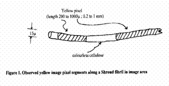
The coloured image pixels have sharply terminated ends along each
fibril; that is, they do not just fade out but have clear cut ends [11]. Any
theory of image formation must explain
these characteristics of the individual image pixels.
Burns and scorch areas on the Shroud
There are burn areas on the Shroud near
the patches from the 1532 fire at
The Wounds and Blood Markings on the Shroud
Unlike the image features, which are
negative, with lights and darks as seen by the eye being reversed in the
photograph, the blood and wound markings photograph just as they are seen by
the eye.
Detailed forensic and medical studies over
decades show that the Man in the Shroud was dead, but that there had been no
decomposition of the body. The blood stains and blood flows are consistent
with those to be expected from a scourging and crucifixion.
The stains on the Shroud from the wounds
have been shown to be human blood of
The principal wound marks are: about 120
scourge marks which match those that would be made by a Roman flagellum; the wrists and feet are
pierced by marks corresponding to the nailing of a crucifixion; there are skin
abrasion wounds along the upper back and shoulders which would match those from
carrying a heavy burden; there is a
circlet of head wounds which would match
those from a crown of thorns; there is a large wound in the side of the chest
which would correspond to that resulting from the thrust of a spear. The blood and serum flows
from the chest wound indicate that the victim was dead when the image on it was
made. All of these wound details correspond to the descriptions of the passion
and death of Christ in the New Testament.
The blood stains from the wounds were not
disturbed by whatever process took place in the formation of the image, nor by
whatever process occurred when the Shroud was separated from the enclosed dead,
un-decomposed body that it had enveloped that is to say, there is no sign of
the blood clots having been disturbed or pulled apart. There is no image on the linen beneath the
blood stains.
There are many other forensic and medical
details that are important, but special treatises should be consulted. (e.g. Barbet, Bucklin, Lavoie et al., Baima Ballone et al., [12,13,14,15]
).
Other substances on the Shroud
Samplings of particulate matter on the
Shroud by Frei [16] have shown the
presence of pollen , spores, bacteria
and mineral dusts.
These are consistent with a conclusion that the Shroud during its
history has been in
On the feet, knees and the nose are found
particles of the travertine aragonite
(limestone) dust which also occurs in the
Traces of
myrrh and aloes have also been found on the
Shroud. These would correspond to the historical account of the Entombment in
the Gospel of John as follows:
John
19:38-41 “ … Joseph of Arimathaea ---
asked Pilate to let him remove the body of Jesus. …and he brought a mixture of
myrrh and aloes weighing about a hundred pounds. They took the body of Jesus
and wrapped it with the spices in linen cloths, following the Jewish burial
custom.”
Images of Roman coin from around 29 A.D. can be seen over the eyes
of the Man in the Shroud
 The
eye areas of the image on the Shroud
show two small, round, button-like objects on which have been found parts
of the Greek inscriptions of the lepton
coins of Pontius Pilate, issued by him in Jerusalem around 29 to 30 A.D.
The presence of these coin-like objects was discovered by Jackson, Jumper et al. of STURP [17]. Shortly thereafter, the
identification of the objects as lepton coins issued by Pilate was made by Fr. Francis Filas S.J, [18,19].
Further verification of the coin findings has been made by M. Moroni [10] and by P.L.
Baima Bollone/N.Balossino [10]. Other
studies of coin images and possible other markings on the Shroud have been made
by A. Whanger [20].
The
eye areas of the image on the Shroud
show two small, round, button-like objects on which have been found parts
of the Greek inscriptions of the lepton
coins of Pontius Pilate, issued by him in Jerusalem around 29 to 30 A.D.
The presence of these coin-like objects was discovered by Jackson, Jumper et al. of STURP [17]. Shortly thereafter, the
identification of the objects as lepton coins issued by Pilate was made by Fr. Francis Filas S.J, [18,19].
Further verification of the coin findings has been made by M. Moroni [10] and by P.L.
Baima Bollone/N.Balossino [10]. Other
studies of coin images and possible other markings on the Shroud have been made
by A. Whanger [20].
The presence of these coin images support
a date of origin for the Shroud image to the era of the crucifixion of Christ.
Their importance was generally
downplayed in the 1980’s, apparently because it was felt by some that their existence would weaken the
pressure then being exerted to permit a carbon-14 dating of the Shroud.
There are hundreds of these lepton coins
of Pontius Pilate extant today. They show a curved astrologers staff or lituus together with the curving
Imperial Greek inscription IOU KAICAPOC,
which is a shortened form of the full title, TIBEPIOU KAICAPOC (‘Tiberias
Caesar’). Sometimes the inscription appears with a C instead of K, for example as CAICAPOC. On the Shroud
photographs Filas discovered the curving letters UCAI of the inscription
together with the lituus. The
markings are more clearly seen on second and third negative off-prints,
apparently because the interfering weave pattern of the linen of the Shroud
becomes progressively suppressed.
 The
physical means by which images of metal
coins could have become imaged on the Shroud, as well as the images from
organic elements such as flesh, hair, and blood is clearly important for any
fundamental explanation of the image mechanism.
The
physical means by which images of metal
coins could have become imaged on the Shroud, as well as the images from
organic elements such as flesh, hair, and blood is clearly important for any
fundamental explanation of the image mechanism.
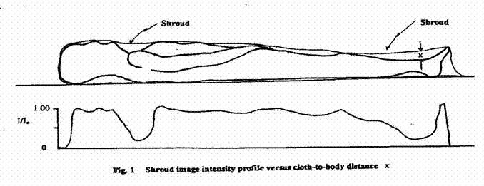
THE THREE-DIMENSIONAL NATURE OF THE BODY IMAGE ON THE SHROUD
It was Paul Vignon [21] who first noted that the darkness or
lightness of the image seemed to be related to how close or how far the
particular image area would have been from a body enveloped in the cloth.
[Figure 1]. This is the famous three
dimensional quality of the image that was firmly established on a
quantitative basis by the STURP scientists, especially by Jackson, Jumper, Ercoline and German. [22, 23, 24]. This then means that the closeness of spacing
of the image elements or pixels on the cloth varies quantitatively with the
distance of the linen Shroud from the enveloped body [ Fig.1].
We shall return to this 3-dimensional
problem when we investigate how the image got onto the cloth in the first place
- in all probability by some sort of radiation which drops off in intensity
with distance from the body by being absorbed or attenuated in the air gap
between body and Shroud.
THE NATURE OF THE SHROUD IMAGE AS EXAMINED BY PHYSICS AND CHEMSTRY
The Image Characteristics
Before we discuss image-transfer theories,
that is, how the image got from the enclosed dead body of The Man in the Shroud
to the cloth itself, let us set out the now well-established chemical
characteristics of the image on the Shroud.
What is the image chemically?
The image consists of separate, tiny,
straw-yellow coloured image elements or image ‘pixels’ along individual linen fibrils. The fibrils
themselves are around 15 to 20 microns in diameter. The coloured pixels range in length along the fibrils from about 200 microns to around 1000
microns.. The mage pixels are abruptly terminated on the fibrils, that is to
say , they have sharp beginning and ending boundaries; they do not fade out
gradually but start and end at full colour.
The pixels occur only on the topmost fibrils of the top linen threads on
the image side of the Shroud, so that the image is extremely superficial on the
linen. The threads and fibrils that are
deeper into the weave are colourless.
Where one image fibril crosses over another fibril the undermost fibril
is colourless. Thus the image mechanism does not penetrate the cloth or the
individual threads. It is a ‘top-of-the-topmost-threads-only’ mechanism.
The intensity of hue of the individual
image pixels is always the same. It is only the closeness or spacing of the
individual pixels that causes the variation in the image shading from light to
dark image areas. Thus the chemical, process is very uniform and abrupt, and
moreover is not continuous along any fibril, but is always quantified into
discrete lengths of 200 -1000 microns.
There is no variation of pixel hue on the underside of the body image where the
weight of the body would be pressing down on the Shroud, as compared to the top
image; thus the individual image
elements or pixels are of the same intensity regardless of the contact pressure
of the body against the cloth as compared with areas where there is no contact
and where there is an air gap between the body and the enveloping Shroud. Since
the individual pixel hue is always the same, regardless of separation or
contact pressure, then clearly the chemical process involved in image formation
is a very precise one.
Linen is a cellulose fibre from the flax
plant. Cellulose is polycellobiose
with the chemical formula (C6H10O5)x
. Its gram-molecular weight is 162.14.
When cellulose ages by being exposed to light
for a long time, it takes on an ivory
colour, or it ‘degrades’. The change occurs because some of the chemical bonds
or molecular linkages are altered by the action of the light. This chemical
degradation of linen can also be caused quickly in the laboratory by applying
heat at around 200 -300 deg C, or by
applying strong acids such as sulfuric acid.. The chemical change is called an acid oxidation/dehydration, and it
produces a repetitive, alternating or
‘conjugated’ double-bond molecular structure –C=O- which causes the straw-yellow colour or chromophore structure
The scientific consensus today is that the image on the Shroud is fundamentally due to a chemical discoloration of
the cellulose fibrils of the linen in each tiny image element or ‘pixel’.
This chemical change is technically called an acid -oxidation of the cellulose
fibrils to produce the straw-yellow chromophore groups –C=O- which color the
individual pixels. While to the eye the colour of the image areas is sepia or
pale brownish, under the microscope the colour in the individual image elements
or pixels is straw-yellow. This chemical process for forming the straw-yellow
colour of the pixels on the fibrils was first established by Heller and Adler [25] ; a recent
summary is by Moran and Fanti [26].
How
did this chemically-produced image get transferred onto the Shroud from the
enveloped dead body in the cloth?
Over the past century many explanations
have been put forward to explain how the mysterious image transferred from the
enveloped dead body to become imprinted
on the Shroud. As scientific data
accumulated, various theories were successively put to the test of their
consistency with all the observed image characteristics, and one by one have
been discarded to leave only two. In
this section these various discarded theories will be presented first, followed
by the two scientific answers to the
problem. The principal image formation theories can be classified as follows:
Fraud
image formation theories:
·
Was the Shroud image made by an artist?
The ‘painting fraud’ theory. The ‘renaissance genius’ fantasy.
·
Was the image made by photographic means?
·
Was the image made by a scorch? The ‘hot
statue fraud’ theory
Scientific
image-formation theories:
·
Was the image made by perspiration and
vapors from spices? The Vignon vaporgraph theory.
·
Was the image made by some sort of
radiation? The thermal degradation of cellulose theory.
It must be kept in mind that any image
formation theory must be judged today,
not only by its ability to explain one, or a few, or many, of the image characteristics, but also
in the light of the great mass of evidence for the Shroud’s authenticity that
has accumulated over a century of
serious, dedicated scholarly and scientific research as outlined above. No
fraud theory can face this weight of evidence; consequently fraud theories
today are of minor interest. They will
be outlined here only for background information.
Fraud image-formation theories
‘The
Shroud image is a painting’ theory
Painting requires paints, that is pigments
of various colours dispersed in a painting medium such as oil, tempera, casein,
water etc. Then, the paint is applied to
the support cloth , canvas, paper etc.
usually by a brush or knife which always leave characteristic marks. The
shadings or values of the painting which give it its depth and realism are made
by applying lighter or darker values of the pigment. Paintings are positive
images, that is, shadows and recesses are darker and prominent features are lighters
in value. The shroud image is a negative, with reversed black and white
values. It could not have been painted
before the knowledge of negative
images became known in the middle of the 19th century when black and
white photography was discovered. Conclusively, there are no paint pigments, no paint medium ( oil, egg tempera,
casein, etc,) nor any brush marks on the Shroud. Therefore the Shroud image is
not a painting of any kind.
The only serious proponent of this
painting forgery theory was the late Walter
McCrone who was reputable in the
field of microscopic chemical identification of minerals, dust particles,
chemicals and other substances found on
objects. He claimed to have found iron
oxide on the Shroud, along with traces of vermilion which is used in paints.
The miniscule vermilion specks are today
considered to be entirely irrelevant, and to have come from inadvertent contamination by various artists who painted
copies of the Shroud over the centuries.
In spite of the fact that all the other
evidence was missing - that is to say, no shading of iron oxide, no paint
medium, no brush marks and so on - McCrone still strenuously proclaimed his view
that the Shroud was a painted hoax.
Heller
and Adler [25] soon
demonstrated that the iron oxide was not outside on the linen fibrils as in
paintings but inside the cellulose
fibrils themselves, and that this was due to the traditional retting process of
making the linen from the flax plant by soaking it for days in ponds or streams where it
chemically absorbs iron, strontium,
calcium, potassium and many other water
soluble minerals into the cellulose pores.
McCrone refused to consider their
evidence and eventually became isolated from serious work on the matter.
His assertions are still circulated by mass media interests, but not in serious
work.
Many professional artists such as Picek [27] have also discussed the
technical incompatibility of various
painting techniques with the Shroud’s
image characteristics. In spite of this, however, presentations are
occasionally still made presenting someone such as Leonardo da Vinci as the ‘genius Renaissance
art forger’ of the Shroud. Such claims
are mere propaganda or sensational exploitation of his reputation. Again, it
must be pointed out that, even if an
actual convincing demonstration that a negative
shroud-like image could be painted, it would still be irrelevant to the
Shroud’s authenticity as the true shroud of Christ, since it would be
contradicted and refuted by the mass of other evidence for authenticity which
it can not explain. Painting forgery
theories of Shroud image formation today are therefore unacceptable.
Was
the Shroud image made by photographic means?
Since the Shroud and its image are now
securely dated historically to the 1300’s in Europe, and to the years from 1204
to 944 in Constantinople, and to Edessa prior to 944, the argument that a negative photograph could have been fraudulently imprinted on cloth many centuries before the invention of photography is not
worth discussion.
Was
the Shroud image made by a scorch from a
heated metal statue?
This theory originated from the
observation that image on the linen has
many of the characteristics in hue and shading that a thermal scorch on
linen can produce. Consequently it was
theorized by Ashe [28] and others
that if a linen cloth were to be draped over a metal effigy heated to 250 deg.
C ( which is about the scorch temperature threshold for linen) then the image
could have been produced. Since the
draped cloth would have various air gap distances between statue and the cloth
draped over it, and since thermal radiation is attenuated in air to some degree
as it passes across the air gap, then some three dimensional information might
be transferred to the cloth, points closest to the statue being more scorched
than those farther away. However,
thermal radiation spreads out widely, and so would not give the required
definition to the image features. In addition, the attenuation rates, or the
drop off in radiation intensity with distance traveled through air, could
not match those observed on the Shroud
image.
Furthermore, a true scorch also causes
linen to fluoresce under ultraviolet or X-ray examination. But the STURP team
of investigators who examined the Shroud
in 1978 found that, although the burnt patches on the Shroud from the
1552 fire did fluoresce as expected, the image areas showed no signs of
fluorescence at all [6,8,9]. Therefore,
the image on the Shroud was not made by a thermal scorch.
Although, since
it is a ‘fraud theory’ , the hot statue theory
is today untenable against the
accumulated evidence for authenticity, it
has been historically very useful
because it proposed a chemical
degradation of the cellulose, and so spurred investigation of an essential
ingredient of any plausible image formation theory.
SCIENTIFIC IMAGE FORMATION THEORIES
General
Today there
are only two serious scientific theories
for chemically altering the Shroud’s cellulose and producing the observed
image. First, there is the complex vaporgraph hypothesis, which involves contact, vapour diffusion, chemical
catalysis, heat and ageing. Apart
from being very involved and complex,
this theory cannot meet many of the
essential image requirements.
Second, there is
the radiation hypothesis which meets
all the image characteristics, is intrinsically simple, but which then raises
very difficult questions with regard to the
origin of the image-forming radiation.
1. Was the Shroud image made by a
vaporgraphic process?
Vignon
[21] presented
this, the first scientific attempt to explain the physical and chemical
processes by which the image might have been imprinted on the Shroud by a
natural process. He theorized that a
body in pain produces perspiration containing urea, and that urea may
chemically change to produce ammonia. Then he reasoned that the embalming
spices myrrh and aloes mentioned in the Gospel of John (
Vignon’s
ingenious theory was soon found to have various untenable features. The
diffusion of vapours could not have
produced the necessary sharpness of definition of the Shroud image. Also,
diffusing vapours would permeate the fibrils, whereas the
Shroud image is only on the topmost fibrils of the inner side of the cloth and does not penetrate into
the linen as would have been the case with a diffusion process.
A variation on the Vignon theory is the latent image hypothesis. Pellicori [6] was able to
experimentally produce chemical alteration of cellulose to closely resemble the
image area discolorations by applying small amounts of essential oils, such
as myrrh and aloes, or olive and other oils, to a linen cloth, and then baking the cloth
at temperatures of around 150 deg.C for several hours. In this way he was able
to produce discolouration on the linen which closely matched the discolouration characteristics of the
Shroud image areas
His
theory, however, does not explain how the image discolourations could be made
to vary so as to match the three dimensional
image feature of the Shroud, nor to reproduce its observed fine optical
resolution. The main value of this theory is again to focus attention on the
chemical alteration of the cellulose as being the source of the image on the
cloth. As a transfer theory it fails because only radiation transfer with its
attenuation in an air gap can fully account
for the three-dimensionality of the image. It also could not produce the
discrete, sharply terminated image pixel lengths that are observed.
2. Radiation as an image transfer and image
formation explanation
 Radiation with
wavelength shorter than 340 nm (3.4 x 10⁻⁹ m), such as X-rays
and energetic ultraviolet radiation, can directly
degrade cellulose. However, radiation of such short wavelength is also so
energetic that it not only chemically produces chromophore rearrangement, but
at the same time also pyrolizes or explosively disrupts the cellulose of the linen and thereby alters its appearance and colour.
This rules out the action of energetic, short wavelength radiation as an
image transfer means, since the Shroud image areas shows only the chemical
alteration of the cellulose to the pale
straw-yellow colour but no fibril damage.
Radiation with
wavelength shorter than 340 nm (3.4 x 10⁻⁹ m), such as X-rays
and energetic ultraviolet radiation, can directly
degrade cellulose. However, radiation of such short wavelength is also so
energetic that it not only chemically produces chromophore rearrangement, but
at the same time also pyrolizes or explosively disrupts the cellulose of the linen and thereby alters its appearance and colour.
This rules out the action of energetic, short wavelength radiation as an
image transfer means, since the Shroud image areas shows only the chemical
alteration of the cellulose to the pale
straw-yellow colour but no fibril damage.
Longer wavelength, that is to say, weaker
radiation, (visible light, infrared,
microwave) does not cause any damage
to linen, but does cause the needed colour
change which forms the observed image. However it is energetically quite weak
and can not directly affect the cloth except over very long times of
exposure. Moreover, radiation would penetrate the threads and fibrils to color
them interiorly which is not observed. Consequently, even long wave radiation, which is is a candidate
because it is energetically suitable, still requires some indirect mechanism of energy absorption which (1) raises the temperature at the
surface of the cellulose fibrils to 200 to
300 C, but not high enough to damage the cellulose or pyrolize it,
and (2) this indirect mechanism would
also have to correctly produce the
observed discrete, separated pixels having the observed lengths of 200 to 1000 microns, and which, moreover, must be abruptly terminated
at each end as mentioned above.
The various types of radiation are listed
in Table 1 together with their effect on cellulose.
Table
1
Properties of Various Radiations and Image Formation Requirements
A. Electromagnetic Radiation
Type Wavelength Energy Attenuation in air Action on linen Remarks
X-ray 1 – 10
nm strong Negligible Damaging Ruled out
UV 10-40 nm moderate “ Damaging Ruled out
Visible 400-800 nm weak “ Yellowing Ruled out
Infrared .001 -0.3 mm weak Variable Yellowing Ruled out except for far-infrared
Microwave 0.3-1 mm weak Matches Shroud data Yellowing O/K if indirect
B. Flux of Elementary
Particles
Proton -- very high Matches some data Yellowing OK for few features
Neutron -- “ Nil attenuation Adds C14 No image effect
1 nm (nanometer) = 10-9 meter
1000 nm = 1 micron (μ) =10-6
meter
1000 microns = 1 millimeter = 10-3 meter
-----------------------------------------------------------------------------------------------------------------------------------------------------------------
To sum up: The image was undoubtedly formed on
the Shroud by a chemical alteration
of the cellulose of the linen. The
energy for this chemical change would have to be supplied in some unknown
manner by some form of radiation emanating from within the body of the Man in
the Shroud. The possible candidate types of radiation are microwave and
far-infrared (Table 1) Power [29,30,31,32]. The
basic, image forming microwave radiation was possibly combined with a flux of proton particles
(Table 1) as has been theoretically and experimentally investigated by Rinaudo [33]. (Neutron particle radiation would also transmit energy and generate a uniform
diffuse straw-yellow colouration, but neutrons
interact very weakly with air molecules
and so do not progressively attenuate across the air gap between the enclosed body and the
cloth, and therefore would not produce
the beam intensity variations needed to generate a three- dimensional image on
the cloth.)
The
requirements for radiation as an image producing process are (1) that it be partially absorbed or
attenuated in passing through the air
gap between body and the enveloping Shroud so as to produce the intensity variations which generate the image on the cloth, (2) it must have sufficient energy to chemically act on
the cellulose ( directly or
indirectly) so as to produce the basic
straw-yellow image pixels, but not enough energy to destructively attack the
cellulose, (3) it must produce the image
pixels in the observed sizes and
with the observed sharp ends or boundaries, and
(4) it must explain the
apparent vertical alignment of the image with the earth’s gravity
1. Attenuation of radiation needed to transfer and form an image
The strength or intensity of the Shroud
image at each point on the cloth was studied intensively in 1977 by the STURP team and again in 1978-82
[17,22,23,24]. Their data have been
analysed by Power [29,30,31,32] and are graphed in Figure 1and 2. below.
The basic attenuation relationship is the
Lambert-Bouguer intensity law
I/I0 m= e-kx
where I0 is
the initial radiation intensity presumably from the enclosed body, I is the
radiation intensity after traveling a distance x across the air gap between the
body and the Shroud, and k is the
‘attenuation constant’ which depends on the nature of the radiation and the
nature of the material through the radiation is traveling. Here we are
concerned only with attenuation in moist air.
The air gap distance
x is shown in Figure 1. The observed Shroud image
intensities, the attenuation of radiation intensity across the air gap distance
x, and
several possible curves to fit
the data are shown in Figure 2.

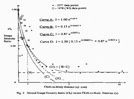
.
We see that greatest intensity image
points on the Shroud ( I/Io nearest to 1) occur at separations (x)
of less than half a centimeter between
cloth and body, for example at the
forehead, tip of the nose, hands and knees.
Lower intensity images ( I/Io < 0.5 ) occur at distances
from 1 cm out to 6-9 cm. such as at the eye sockets, neck and ankles.
The drop off in intensity with increasing
separation of cloth from the body is
called attenuation. Now since the attenuation rates in air for various
types of radiation are well known, this allows us to select the type of
radiation which best fits the observed attenuation in intensity of the Shroud
image. It turns out that radiation of
wavelength of 0.3 to about 1 millimeter has the required attenuation rate in
air ( k value around 0.855); this is
mainly in the microwave region (Curve C).
A second component of radiation, having very little attenuation with
distance ( k = 0.00023), and which may be a
subsidiary proton/neutron flux as proposed and experimentally evaluated
by Rinaudo [33], is also evident (
Curve B). The ‘curve of best fit’ is
thus a two- component radiation Curve D. (Curve B + C = Curve
D)
2. The energy necessary for radiation to produce the Shroud image
Microwave radiation, because it has the
required attenuation in air, could transfer image information by an attenuated intensity flow from body to cloth.
But, does microwave radiation also have sufficient energy to cause the necessary chemical alteration of cellulose to
form the image?
Power
[32] estimated the energy
required to degrade cellulose thermally
and found it to be about 286 Joules of energy per gram of linen
fibrils chemically altered to form the molecular chromophores –C = O- that make up the
straw-yellow image pixels. This
relatively small energy is well within the range of energies of a microwave
flow.
3.
Formation of the image pixels by indirect energy absorption
However, this small amount of energy must
also be able to couple to the cellulose in such a manner as not only to produce
the straw-yellow colour, but to do so in the observed discrete sizes of the
image ‘pixels’, and also to affect only the topmost fibrils
on the topmost threads of the linen. This combination of requirements is quite
demanding for any theory.
.
To repeat, in addition to fitting the
attenuation requirement and having the necessary energy, a candidate type of radiation must be able
(1) to act chemically on the linen and
produce the pixels of the yellow colouring, with no explosive or pyrolitic damage, and 2) the
pixels produced by the radiation on the fibrils must be 200 to 1000 microns
long and abruptly terminated at each end.
No type of radiation can do all this by direct absorption, since any radiation
will penetrate to some extent into the threads and discolour the inner fibrils,
and this is not observed. However,
microwave radiation because of its ability
to heat water efficiently can meet
the requirements by being
absorbed, not directly by the linen, but by being absorbed by micro-droplets of water condensed
onto micro-crystals of various common
mineral salts which are know to be present on the Shroud. The microwave radiation heats
the micro-droplets of water to form superheated steam droplets which then
catalyzes the necessary chemical change in the cellulose to form the individual straw-yellow image pixels in the
proper size range, Power
(35).
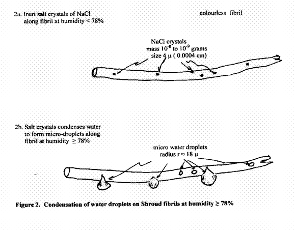
Figure 2. depicts how high humidity (
>78%)- such as would necessarily prevail initially in the air gap between
the Shroud and the enveloped dead body-
would automatically form condensed micro-sized water droplets onto
micro-crystals of mineral salts known to be present in copious amounts on the
Shroud [26].
4.
Formation of image pixels in the proper observed sizes
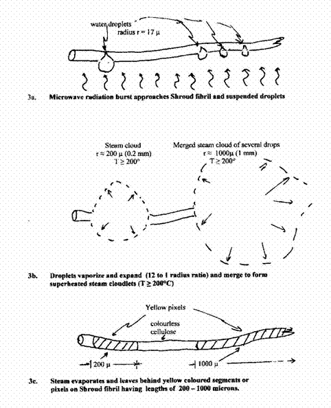
Microwave radiation impinging on the
micro-droplets will heat them and form superheated steam cloudlets with temperatures of above 200 degrees, which is
the minimum temperature needed to quickly degrade cellulose to form the yellow
pixels. Superheated steam immediately causes the formation of the chromophore
bond ( -C=O-) to produce the straw-yellow
spots or ‘image pixels’ on the cellulose fibrils, in the proper observed
lengths ( 200 to 1000 microns ; 0.2 to 1 mm),
and with their observed abrupt ends at the boundary of each separate steam cloudlet (Fig. 3), R. N. Rogers [34] and Power [35].
Conclusions from the Scientific Facts as to the nature of the image
on the Shroud and the means of image transfer to the cloth from the enveloped
Body of the Man in the Shroud.
1.
The image on the Shroud is a result of a chemical alteration of the cellulose
of the linen cloth brought about by a
natural energetic means and not by any human action or forgery. The specific chemical reaction is an
acid, dehydrative oxidation of the cellulose to form conjugated –C=O-
structures which produce the observed straw- yellow colour of the image
elements or ‘pixels’.
2. The natural energetic means that brought
about the chemical alteration, and the image transfer means that carried the
three-dimensional image from the body to the linen cloth, are consistent with a
predominantly low-energy radiation in the microwave region at a
wavelength of 0.3 to 1 millimeter (Power
[35]. Supplemenetary proton/
neutron radiation proposed by Rinaudo [33] may also be involved in the formation of some
subsidiary features of the Shroud image,
such as the image of Roman coins over the eyes, and the faint X-ray character
of some body features of the fingers and the jaw, and possibly in some minor
enhancement of the carbon-14 content of the cloth.
3.
The Scientific Facts together with the Historical Facts point overwhelmingly to
the authenticity of the Holy Shroud of
4.
The theory of the image
transfer process by some sort of radiation is presently still at the lower level of proof of
‘preponderance of the evidence’.
5.
There is currently no scientific consensus at all on the precise nature and source of the image-transferring radiation.
The next step is to try and formulate a
physical theory which can reasonably (and preferably eventually
experimentally) demonstrate the nature
and source of the radiation which transferred the energy which imprinted the observed image of
Christ on the Shroud.
Where
did the (microwave) radiation that formed the image on the Shroud come from?
 The
central problem here is this: To form the observed image on the Shroud, the present
scientific evidence indicates that radiation would have had to emanate from
within the dead body of the Man in the Shroud.
But, radiation does not come from dead bodies. Therefore, such image forming radiation, if
it occurred, must have been due to been an extraordinary event.
The
central problem here is this: To form the observed image on the Shroud, the present
scientific evidence indicates that radiation would have had to emanate from
within the dead body of the Man in the Shroud.
But, radiation does not come from dead bodies. Therefore, such image forming radiation, if
it occurred, must have been due to been an extraordinary event.
Some researchers on the Shroud who believe
that the image was formed in connection
with the Resurrection of Christ have
suggested that some ‘burst of radiation’ occurred [36], but no plausible
physically based explanation for such an
extraordinary radiation event has been put forward. Most scientific researchers, indeed, have
preferred to keep away from any ultimate
aspect of the problem as long as
possible and to concentrate on the details of the image which can be studied
scientifically.
Our position here is that any ultimate question for science may still
be premature. This is because there now
exist several new scientific approaches which it appears may eventually offer a
scientific explanation, although not an ultimate reason of course, for the
extraordinary generation of the necessary image-forming radiation.
New scientific approaches needed?
These new approaches are (a) various variable speed of light (VSL) theories [37-42], and (b) the exploration of the application of negative
pressure-energy flow models to
astrophysical problems.[46].
Among the VSL approaches is that of compressible energy flows, which
already offers tentative explanations for the
weak energy release required to form the observed Shroud image [40,41,42]
This latter theory is an application of standard gas dynamics
and aerodynamics theory [43,44], and as such it is based on a century of scientific and engineering results. It
introduces the VSL condition as an integral, physically-based formulation.
The compressible energy flow
equation is
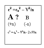
c2 = co2
– V2/n
(1)
where c is the variable speed of light, co
is the constant or static speed of light in space ( 3 x 10⁸ m/s) in the absence of
any relative flow V, and n is the
number of ways the energy of the compressible flow is divided. The flow is
understood to be for unit mass, so that mass does not appear explicitly. In
this equation the wave speed c decreases from the maximum static wave speed value co as V
increases and reaches zero at a maximum
flow escape speed to a vacuum, Vmax = n1/2 co. This
maximum flow speed is superluminal in all cases for n greater than 1.
In highly accelerated elementary particle
flows, n approaches 1, and the compressible energy flow equation then yields
the Lorentz-Fitzgerald ‘contraction
factor’ of special relativity theory on a purely physical basis, since,
rewriting Eqn. 1 with n = 1, we
have
c/co = [1 – (V/co)2]1/2
where the bracketed expression is the
familiar contraction factor.
If the flow perturbation is a pulse, then
we have
c2
= co2 – V2/n – 2cV/n.
In all known material gases n is a
positive integer, but the possibility of it being, alternatively, a negative
number, either integral or fractional,
raises the possibility of a binary, evolving universe encompassing both
our ordinary, condensed energy matter and the astronomical ‘dark matter’.
The adiabatic equation of state in compressible flow is
pvk = const.
Where n
= 2/(k-1) ; k = (n+2)/n = γ = cp/cv ,the ratio of
specific heats of the fluid. As in the
energy equation, n and k can be either
positive or negative. Positive n values correspond to all know material gases
where compression waves are the rule and wave speeds are equal to or less than
the static wave speed co
(World A). Negative values of n (World
B) yield hypothetical exotic fluids
where k can be variously positive or negative. Rarefaction waves are the rule.
Here wave speeds are always superluminal
, that is c is greater than the static wave speed co.
In 1999 the discovery of anomalous
redshifts in the cosmic microwave
background CMB by Bachall, Ostriker,
Perlmutter and Steinhardt [45] was interpreted to mean an accelerating
expansion of the universe under the influence of an anomalous ‘negative
pressure energy’, which was called ‘
dark energy’.
The proposal of Kamenshchick, Moschella and Pasquier [46,] that the dark matter and dark energy can both be related to the so-called Chaplygin gas, also called the
‘tangent gas’ in aerodynamic theory [43, 44], has now opened the door to
widespread active investigation of the suggested involvement of negative-n
states with dark matter and dark
energy.
In the Chaplygin gas n and k are both
negative and equal to -1; its wave speed c,
interpreted as the speed of light, is always superluminal, since in (Eqn
1), with n negative, V2 always adds to the static wave energy co2.
The Chaplygin gas has the following form:
pv-k = const. where k = -1 and n = -1, that is
pv-1 = p(1/v) = const. and
since 1/v = ρ , the density, then we have
p= A/ρ
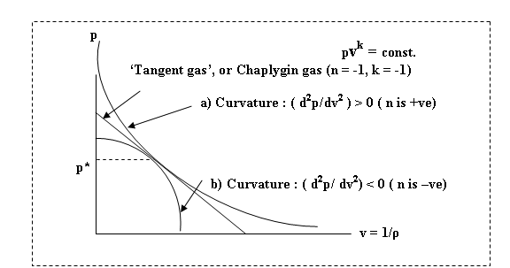
World A ( positive n) and World B (
negative n) Compressible Fluid
Equations of
State
Since the Chaplygin gas curve has negative n (unlike the asymptotic World
A isentropes ( a) where n is positive)
it intersects the v axis.
Therefore, negative pressures can be experienced, and it is this property that
is used to explain the observed anomalous red-shift currently interpreted as
being an accelerated expansion of the
universe [46]. At the present time extensive investigation is continuing into
this new possibility [ e.g. 47,48].
This new World B physical and
astrophysical theory will be outlined in
future Updates to the Website in the context of its application to the problem
of image forming radiation for the Holy Shroud .
References
1 Ian Wilson, The
Shroud of Turin. Doubleday & Company, Inc. Garden City, N.Y.,1978
2. Mechthild Flury-Lemburg, Sindone
2002 . L’intervento conservatio. ( The Shroud 2002. Intervention for
Conservation). Publishing House for the Diocese of Turin (ODPF) 2003. (www.sindone.org/).
3. A. Timossi, La S. Sindone nella sua Contituzione Tessile.
Torino, 1933.
4. Proceedings of the 1977
United States Conference of Research on the Shroud of Turin. March
23-24, 1977. Albuquerque, New Mexico.
Published by: The Holy Shroud Guild, 294
East 150th Street, Bronx, N.Y., 10451 (http://www.shroud.org/).
5. Eric Jumper and Robert W. Mottern,
Scientific Investigation of the Shroud of Turin. J. Applied Optics. 19,
12, p 1909, June 15, 1980.
6. S.F. Pellicori, Spectral Properties of the Shroud of Turin, J. Applied Optics. 19, 12, p 1913, June 15,
1980.
7. J.S. Acetta and J. Stephen
Baumgart, Infrared reflectance
spectroscopy and thermographic investigations of The Shroud of Turin. J. Applied Optics. 19, 12, p 1921, June 15,
1980.
8. Roger Gilbert Jr., and Marion M
Gilbert, Ultraviolet-visible reflectance
and fluorescence spectra of the the Shroud of Turin. J. Applied Optics. 19,
12, p 1930, June 15, 1980.
9. R.A. Morris, L.A. Schwalbe and J.R.
London, X-Ray Fluorescence Investigation of the Shroud of Turin. X-ray Spectrophotometry, 9, 2,
p. 40, 1980.
10. Gino Moretto, The
Shroud: A Guide ( English transl.).
Paulist Press, New York, N.Y. 1996.
11. Kevin E. Moran, Optically Terminated Image Pixels Observed on
Frei 1978 Samples. Intnl. Research
Conference “Multi-disciplinary
Investigation of an Enigma”, Richmond, Virginia, 18-20 June, 1999.
12. Pierre
Barbet, La Passion et la Mort de N.S. Jesus-Christ Selon le Chirugien. Paris.
Dillon & Co. 1950.
----------------------, A
Doctor at Calvary. Image Books, N.Y. 1963.
13. Robert Bucklin, M.D, The Medical Aspects of the Crucifixion of
Christ. Sindon, 7, 1961, pp.5-11.
14. Gilbert R. Lavoie, B. Lavoie, V.J. Donovan, and L.S.
Ballas, Blood on the Shroud of Turin. Shroud Spectrum Intnl. 8, 1983, pp 2-10. See also www.shroudofjesus.com
15. Pier Baima Bollone, M. Jorio, and A.L.
Massaro, La demonstrazione della prezensa
di trace di sangue umano sulla Sindone. Sindon 30, 1981, pp 5-8.
16. Max Frei, Nine years of palynological studies on the
Shroud. Shroud Spectrum Intnl. 3,
1982, pp 3-7.
17. John P. Jackson, Eric J. Jumper, Wm. Mottern, and Kenneth E.
Stevenson, The Three-dimensional Image
on Jesus’ Burial Cloth. 1977 United States Conference of Research on
the Shroud of Turin. March 23-24
1977. Available from Holy Shroud Guild. 294 East 150th Street, Bronx, New York, 10451. See also www.shroud.org/
18. Francis Filas, S.J., The dating of the
Shroud of Turin from coins of Pontius
Pilate. 1982. Cogan Productions, A
division of ACTA Foundation, 11134 Youngstown Ave., Youngstown, Arizona 85363.
19.---------------, What are the Facts
about the Shroud of Turin?. Messenger,
91, 4, April 1981 pp. 12-16. ( Canadian Messenger. 661 Greenwood Ave. Toronto, ON, M4J 4B3).
20. A. Whanger, ‘Polarized Overlay
Technique: A New Image Comparison Method and its Applications, Applied Optics 24,
16, 15 March, 1985, pp 766-72. See also www.duke/~adw2/shroud
21.
Paul Vignon, The Shroud of
Christ. Constable, London. 1902.
22 .John P. Jackson, E.J. Jumper and W. Ercoline. The Three-dimensional
Characteristics of the Shroud Image. Proceedings of the 1982 IEEE International
Conference on Cybernetics and Society. Seattle. Oct. 28-30 1982.
23.------------ Correlation of image
intensity on the Turin Shroud with the 3-D structure of a human body shape. Applied Optics 23
July 1984, pp. 2244-70.
24. John D. German, Jr, . An Electronic
Technique for Constructing an Accurate Three Dimensional Shroud Image. Proceedings
1977 United States Conference of Research on the Shroud of Turin. March
23-24 1977. pp. 234-240. Available from Holy Shroud Guild. 293 East 150 Street,
Bronx, New York. See also www.shroud.org/
25. John H. Heller and Alan D. Adler, A Chemical Investigation of the Shroud of
Turin. Can . Soc.. Forensic Sci. J. 14, 3 pp-81- 103, 1981.
26 Kevin Moran and Giulio Fanti, Does the
Shroud body image show any physical evidence of Resurrection? IV Symposium Scientifique International du
CIELT. Paris, 25-26 Avril 2002.
27. Isabel Piczek, Why the Shroud of Turin
could not have been the work of a clever artist. Los Angeles, Private
publication, 1989.
---------------------, The Shroud of Turin
according to the Professional Arts: Why it cannot be a painting. Private
publication, 1991.
28. Geoffrey Ashe, What Sort of Picture? Sindon pp. 15-19, 1966.
29. Bernard A. Power, Il Meccanismo di Formazione dell’Immagine
dela Sindon di Torino, Collegamento pro
Sindone, Roma, Maggio-Giugno, pp
13-28, 1997.
30.------------------------,
Caratterizzazione di una Lunghezza d’Onda per la Radiazione che Potrebe aver
Creato I’Immagine Della Sindone di Torino. Collegamento
pro Sindone, Roma. Novembre-Decembre, pp. 26-36, 1999.
31. -----------------------, An Unexpected
Consequence of Radiation Theories of Image formation for the Shroud of Turin. Proc. Worldwide Congress Sindone 2000, Orvieto, Italy, Aug. 27-29, 2000.
32. -----------------------, Image
Formation on the Holy Shroud of Turin by Attenuation of Radiation in Air. Collegamento
pro Sindone website (www.shroud.it/) March 2002.
34. R.N Rogers, The Chemistry of Autocatalytic Processes in
the Context of the Shroud of Turin. (www.shroud.com)
35. Bernard A. Power, How Microwave Radiation Could Have
Formed the Observed Images on The Holy Shroud of Turin. Collegamento [ro Sindone Website, Jan. 2003. (www.shroud.it/)
36. Kenneth E. Stevenson and Gary H.
Habermas, Verdict on the Shroud, Servant Boks, Ann Arbor , Michigan, 1981.
37.
John D. Barrow, and J. Magueijo, Phys. Rev. Lett. B 443, 104-110, 1998.
38.
Donam Youm, Variable–speed-of-light cosmology and second law of
thermodynamics. Physical Review D., 66, 043506, 15 Aug. 2002.
39. John D. Barrow, Variations of alpha in
space and time. Phys. Rev. D. 043515,
15Aug. 2002.
40. Bernard A. Power, Shock Waves in a Photon Gas. Contr. Paper No. 203, American Association
for the Advancement of Science, Ann. Meeting, Toronto, Jan. 1981.
41. -----------------------, Unification
of Forces and Particle Production at an Oblique Radiation Shock Front. Contr. Paper N0. 462. American
Association for the Advancement of
Science, Ann. Meeting, Washington,
D.C., Jan 1982.
42.
----------------------, Baryon Mass-ratios and Degrees of Freedom in a
Compressible Radiation Flow. Contr. Paper No. 505. American Association
for the Advancement of Science, Annual Meeting, Detroit, May 1983.
43. A. H. Shapiro, The Dynamics and Thermodynamics of Compressible Fluid Flow. 2 Vols.
John Wiley and Sons, New York, 1953.
44. R. Courant and K. O. Friedrichs, Supersonic
Flow and Shock Waves. Interscience , New York, 1948.
45. N.A. Bachall, J.P. Ostriker, S
Perlmutter and P.J. Steinhardt, Science, 284, 1481, 1999.
46. A. Kamenshchick, U. Moschella and V.
Pasquier, An alternative to
quintessence. Phys Lett. B 511, 265, 2001.
47. N. Bilic, G.B. Tupper and
R.D.Viollier. Unification of Dark Matter
and Dark Energy: the Inhomogeneous Chaplygin Gas. Astrophysics,
astro-ph/0111325, 2002.
48. P.P. Avelino, L.M.G. Beca, J.P.M de
Carvalho, C.J.A.P. Martins and P.Pinto. Alternatives to quintessence model
building. Phys. Rev. D.67 023511, 2003.
![]() MAIN PAGE / The Historical Facts/ The Scientific Facts/ Other Shroud Sites/ Carbon 14 dating
MAIN PAGE / The Historical Facts/ The Scientific Facts/ Other Shroud Sites/ Carbon 14 dating
Copyright © 2003/2004
Bernard A. Power